Mitochondria and Bioenergetics in Health and Disease: It’s Not Just a Power Failure
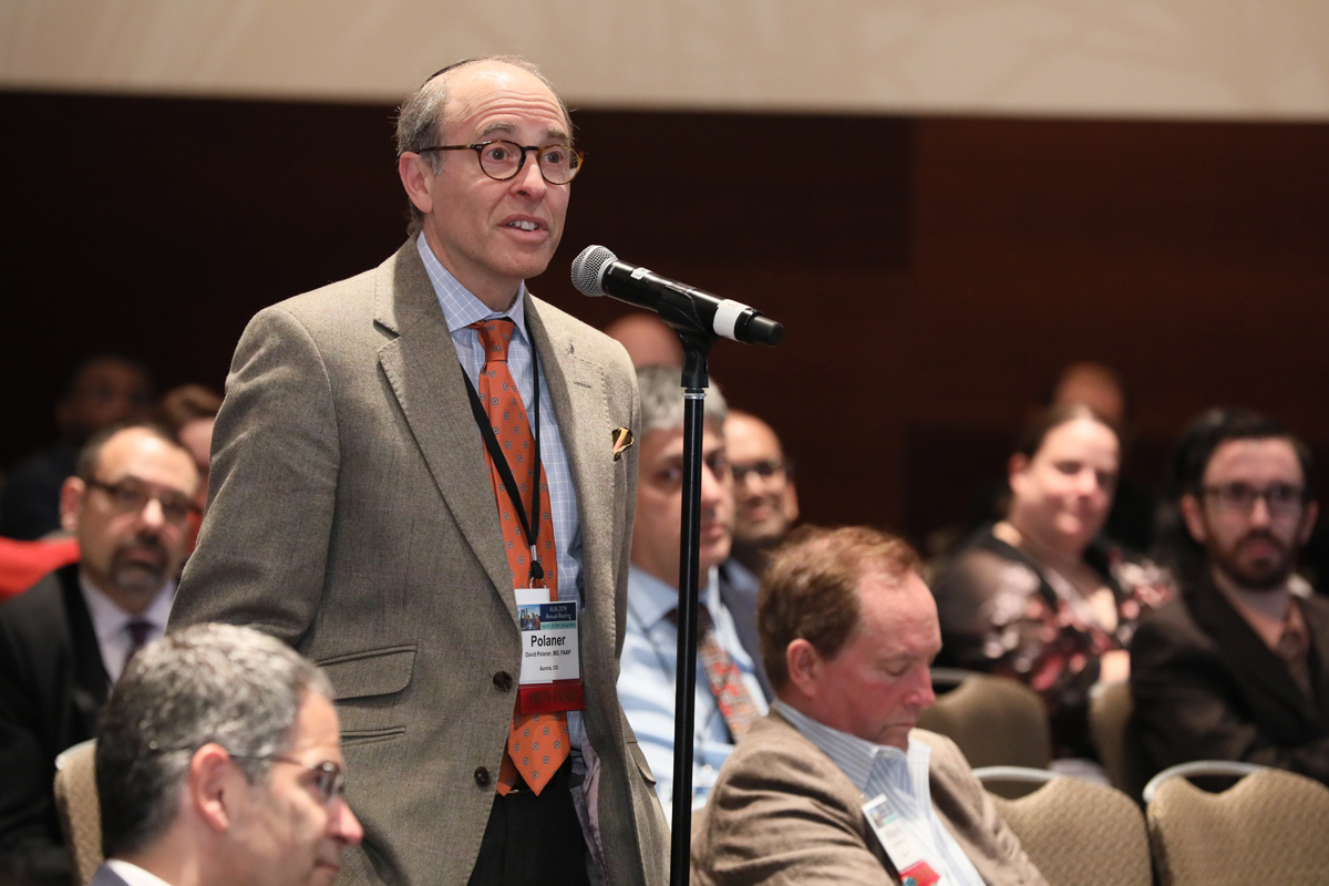
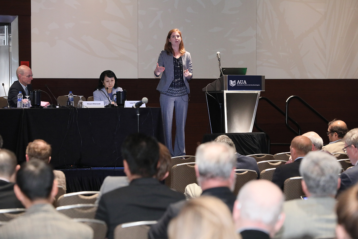
The 65th AUA Annual Meeting was recently held in Chicago, IL. For the third year, we celebrated the successful alignment of the AUA and the IARS in furthering the science of anesthesiology and perioperative medicine by holding a joint AUA-IARS symposium. This year, the topic was “Mitochondria and Bioenergetics in Health and Disease: It’s Not Just a Power Failure”.
Originally derived from ancient aerobic bacteria, mitochondria have their own maternally-inherited DNA (mtDNA) and transcription/translational machineries. The mitochondrial outer membrane is in contact with the cellular cytosol, while the highly folded inner membrane contains oxidative phosphorylation enzyme complexes and the mitochondrial matrix within. Well-known for meeting cellular energy demands via ATP synthesis in the process of metabolizing carbohydrates and fatty acids using oxygen, mitochondria rapidly increase energy production under conditions of cellular stress (spare or bioenergetic respiratory capacity; critical for long-term cell survival). One essential aspect of mitochondrial respiration is generation of reactive oxygen (ROS) that serve physiological functions but are detrimental in excess. Altered metabolism and increased ROS adversely affects other organelles and disrupts cellular homeostasis, requiring defense measures such as the antioxidant enzymes superoxide dismutase, peroxidases and catalase. Mitochondrial ROS can act as signal transducers to trigger expression and/or release of pro-inflammatory cytokines, activate signaling pathways, modulate transcription factors important in redox homeostasis, proliferation/survival, responses to inflammation, protein production, and overall bioenergetics. Importantly, inflammatory mediators and ROS can in turn modulate mitochondrial structure and function, creating a dysfunctional cycle to promote disease pathophysiology.
As the AUA membership realizes, there is now increasing recognition of the relevance of both respiratory and non-respiratory mitochondrial function in a variety of diseases relevant to perioperative care. Prominent examples include: 1) central and peripheral nervous system responses to insults such as hypoxia, ischemia/reperfusion, and volatile anesthetics; 2) cardiopulmonary dysfunction in the context of altered oxygen, ischemia/reperfusion, infection and inflammation; 3) gastrointestinal, hepatic, and renal dysfunction with hypoperfusion and sepsis; 4) neuromuscular responses to anesthesia in the context of intrinsic mitochondrial disease; 5) the effect of altered metabolism associated with diabetes, cardiovascular disease and other chronic conditions that are modulated by medications; 6) organ dysfunction with smoking and environmental insults. While it is likely that the mechanisms underlying these many pathophysiological processes vary, better understanding of how mitochondrial pathways and altered bioenergetics contribute to disease manifestation will be critical for both targeted therapies as well as insights into how to modify perioperative management in individualized vs. population contexts.
The overall goal of this year’s joint AUA-IARS symposium was to highlight important questions relating to mitochondria and bioenergetics that impact upon the perioperative environment. The AUA and IARS are optimal platforms to present these timely topics given that our attendees represent established and emerging leaders in research, education and practice across the bench science to clinical application spectrum. The topic of mitochondria and bioenergetics was also seen as particularly relevant and timely as a focus of the AUA/IARS symposium given that A) although inherent mitochondrial diseases are rare, they have huge perioperative implications, particularly in pediatric anesthesia; B) there is substantial research and clinical focus on the effect of perioperative interventions (including volatile anesthetics) on postoperative outcomes where mitochondria could be a target; C) novel approaches have facilitated focused and nuanced study of mitochondrial structure and function; D) a multitude of medications used by patients for comorbid conditions have the potential to influence the mitochondria and bioenergetics; E) there are now novel therapies targeting mitochondria and metabolism that the perioperative physician should be aware of. Thus, from a research perspective, given many of our colleagues perform studies on mechanisms of disease and/or anesthetic/perioperative drug effects on multiple organ systems of relevance (brain, heart, lung liver, kidney, gut), understanding the roles of altered mitochondrial structure and function as well as bioenergetics mechanisms would help us in improving and individualizing our treatment of patients. Furthermore, given our specialty’s focus on patient safety, understanding unintended consequences of our drugs or other interventions, or the medications taken by patients for co-morbid conditions in the context of mitochondria and bioenergetics also becomes relevant.
The Symposium was moderated by me. The speakers (also serving as panelists during the discussion phase) were Douglas Wallace, PhD (University of Pennsylvania), Elizabeth Jonas, MD (Yale University), Douglas L. Rothman, PhD (Yale University) and Paul Spencer Brookes, PhD (University of Rochester).
Elizabeth L . Whitlock, MD, MSc, Clinical Instructor and T32 Fellow, Department of Anesthesia and Perioperative Care, University of California, San Francisco School of Medicine, San Francisco, California; Co-President, eSAS
Dr. Wallace initiated the symposium with a simply superb presentation on mitochondria through evolution, and the contribution of mitochondrial genes to health and disease. Dr. Wallace (a member of the National Academy of Sciences) is a leader in mitochondrial genetics and one of the most highly cited researchers in this area. He enlightened us regarding how mtDNA plays a role in evolution, normal health, and a range of diseases. He showed us how mitochondrial genes can contribute to normal physiological adaptation in different environs across the world. He delineated how specific mitochondrial genes, and variants within can help explain why certain ethnic populations or inhabitants of regions in extreme climate conditions eat what they do and can tolerate activity (or inactivity) without adverse effects. He then went on to explain how specific mitochondrial genes and variants can contribute to neuropsychiatric disorders. Furthermore, he explained how mitochondrial genes and variants can contribute to volatile anesthetic sensitivity and thus exacerbate cellular metabolic dysfunction in children.
Continuing along the lines of mitochondria in the brain, Dr. Jonas presented her ongoing molecular and cellular work in this area. A neuroscientist/neurologist by training, Dr. Jonas works on mitochondrial metabolism in the brain in the context of stroke neurodegenerative disease, and developmental brain disorders. She focused particularly on the mitochondrial permeability transition pore and the accumulation of a fragment of the anti-apoptotic mitochondrial protein Bcl-xL (deltaN-Bcl-xL) and their contributions to neuronal injury in the context excitotoxic stimulation such as in seizures or in stroke. She further elucidated the potential contribution of mitochondrial mechanisms in anesthetic induced protection.
While cellular and isolated mitochondrial models have provided much insights into non-canonical roles for mitochondria, visualizing their contributions in vivo is a much bigger challenge. Here, Dr. Rothman is a biomedical engineer and expert in biomedical imaging, with development of MR spectroscopy and MRI methods to image metabolic and neurotransmitter pathways non-invasively in humans and in animal models. Metabolism is central to neuroimaging because it can reveal pathways by which neuronal and glial cells use nutrients to fuel their growth and function. Dr. Rothman provided examples of advanced MRI and MRS methods that measure rates of oxygen use and ATP synthesis inside mitochondria (17O-MRS and 31P-MRS), and 19F-MRS that enables measurement of cytosolic glucose metabolism. Calibrated fMRI that uses contrast generated by deoxyhemoglobin, provides maps of oxygen use that track neuronal firing across brain regions. 13C-MRS was shown as the only noninvasive method of measuring both glutamatergic neurotransmission and cell-specific energetics. He introduced novel MRI contrasts to measure brain pH. Overall, these MR methods highlight our emerging ability to assess brain metabolism to better understand brain disorders and guide diagnosis and treatment.
In the last talk, Dr. Brookes elucidated how mitochondrial and their signaling pathways can contribute to anesthetic preconditions in the heart. He demonstrated how a variety of model systems (isolated heart mitochondria, Langendorff perfused mouse hearts, isolated adult mouse cardiomyocytes, in-vivo mouse coronary artery occlusion, cardiomyocyte cell culture) could be used to investigate mitochondrial function (respiration, membrane potential, ROS and NO). He focused on the role of mitochondrial K channels in cardioprotection and showed how volatile anesthetics protect the heart against ischemia/reperfusion injury in anesthetic preconditioning with a particular role for the Slo2.1 channel. Slo2 channels also normally regulate mitochondrial metabolism and could play a role in other organ systems such as brain (and thus be protective in the context of excitotoxic injury).
These exciting talks were followed by robust and healthy discussion among the panelists and the audience regarding 1) How to better understand the heterogeneity of mitochondrial structure and function in normal health (including across the age spectrum); 2) How best to integrate the subcellular/molecular data for in vivo application in the context of perioperative medicine. 3) Providing education and training for clinicians in better appreciation of the roles of mitochondria relevant to clinical practice.
Author

Categories
- Announcements
- Annual Meeting
- ASA News
- AUA: Then and Now
- Call for Articles
- Clinical Research Consortium
- Communications & Website Committee
- DEI
- EAB Report
- Featured Articles
- In Memoriam
- LAB Report
- Learning from Setbacks
- MEB
- Member News
- Mentoring
- Nominations
- President's Message
- SAB Report
- Scholars’ Program
- Statements
- Strategic Planning
- Submission Guidelines
- Wellness
Current Newsletter
Past Newsletters
News & Media


AUA’s EAB and LAB hosted the inaugural presentation of three new awards at AUA’s 2022 Virtual Annual Meeting. Meet the winners!
Publications of Interest
Fiftieth Anniversary of Uncovering the Tuskegee Syphilis Study: The Story and Timeless Lessons
Martin J. Tobin 1,2
1-Division of Pulmonary and Critical Care Medicine, Hines Veterans Affairs Hospital, Hines, Illinois; and -Stritch School of Medicine, Loyola University of Chicago, Maywood, Illinois
Improving Representation in Clinical Trials and Research: Building Research Equity for Women and Underrepresented Groups (2022) (National Academies of Medicine)
Kirsten Bibbins-Domingo and Alex Helman, Editors